25 cells show mitotic spindle defects Early mitotic spindle Biology Diagrams
Blog25 cells show mitotic spindle defects Early mitotic spindle Biology Diagrams Common mitotic spindle defects. Monopolar spindle induced by kinesin-5 inhibition (10 μm STLC, 90 min) in hTERT RPE-1 cell (a). Prometaphase hTERT RPE-1 cell (same cell as Fig. 1b) with spindle axis oriented perpendicularly to the substrate (b). Images of the two focal planes in which the spindle poles resided are shown in the MT column. A 2021 study in The Journal of Cell Biology demonstrated that defects in motor protein function can cause mitotic arrest or chromosome lagging, increasing the risk of genomic instability. Pharmacological inhibitors targeting kinesin-5, such as ispinesib, are being explored as potential cancer therapies due to their ability to disrupt spindle

Correct alignment of the mitotic spindle during cell division is crucial for cell fate determination, tissue organization, and development. Mutations causing brain diseases and cancer in humans and mice have been associated with spindle orientation defects.
Mitotic Spindle: The Intricate Machinery of Cell Division Biology Diagrams
A major contributor to this coordination is the mitotic spindle checkpoint. As detailed in another contribution in this series, defects in mitotic spindle assembly and chromosome alignment activate the spindle checkpoint, which delays cells in M phase. Optimally this delay allows the recovery of the normal spindle and balanced chromosome

Abnormal mitotic spindle geometry: permanent vs. transient. The bipolar geometry of the mitotic spindle is essential for accurate chromosome segregation, and already a century ago Theodor Boveri postulated that supernumerary centrosomes could lead to the production of aneuploid cells [23,24].Observations of mitosis in both transformed and non-transformed cells reveal that multipolar mitotic
PDF A guide to classifying mitotic stages and mitotic defects in ... Biology Diagrams
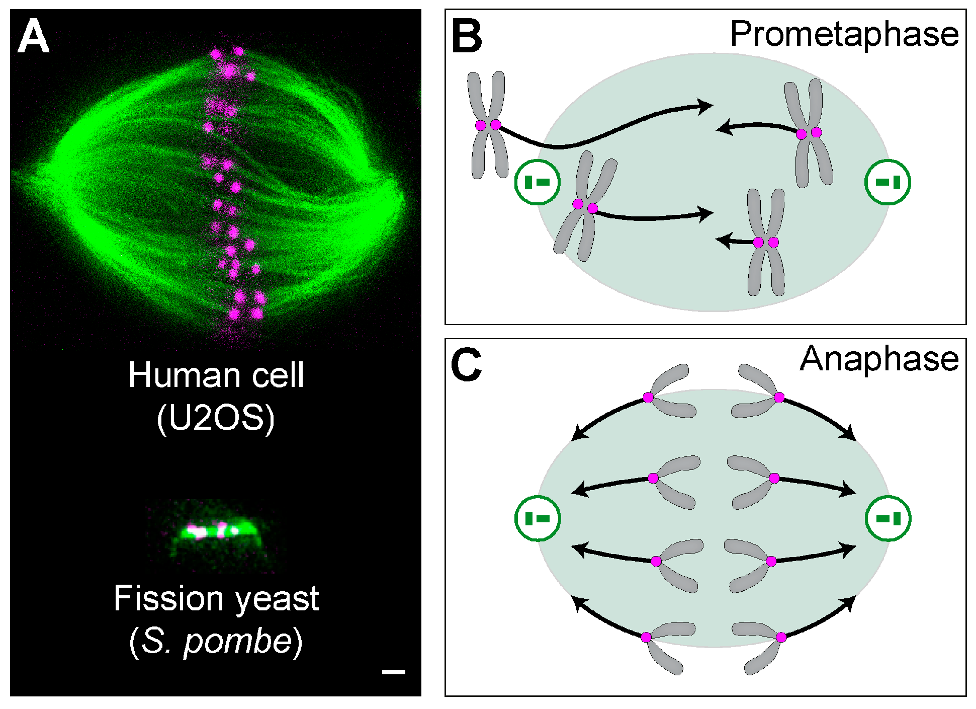
In this article, we provide guidelines for identifying specific mitotic stages and for classifying normal and deviant mitotic phenotypes. We hope this will clarify confusion about how certain defects are classified and help investigators avoid misnomers, misclassification, and/or misinterpretation, thus leading to a unified and standardized In general, lagging chromosomes could be generated by various mechanisms; for example, merotelic attachments (where a single kinetochore attaches to microtubules from opposite spindle poles) would generate lagging chromosomes involving single chromatids, 37 while defects in either sister chromatid resolution or removal of sister chromatid
