Human Skeletal Structure Photograph by Sebastian Kaulitzki Biology Diagrams
BlogHuman Skeletal Structure Photograph by Sebastian Kaulitzki Biology Diagrams Learn about the skeletal system, which provides support, protection, movement and blood cell production for the body. Find out the anatomy and functions of bones, joints, cartilages and bone marrow in humans and other animals.
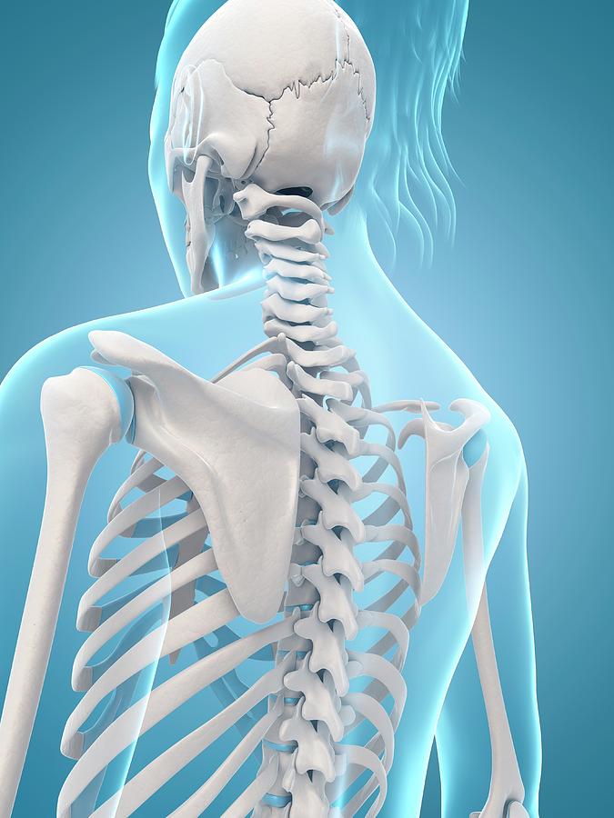
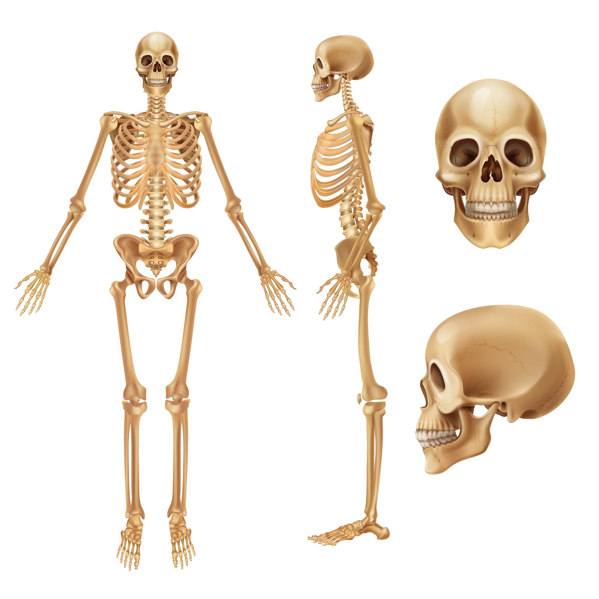
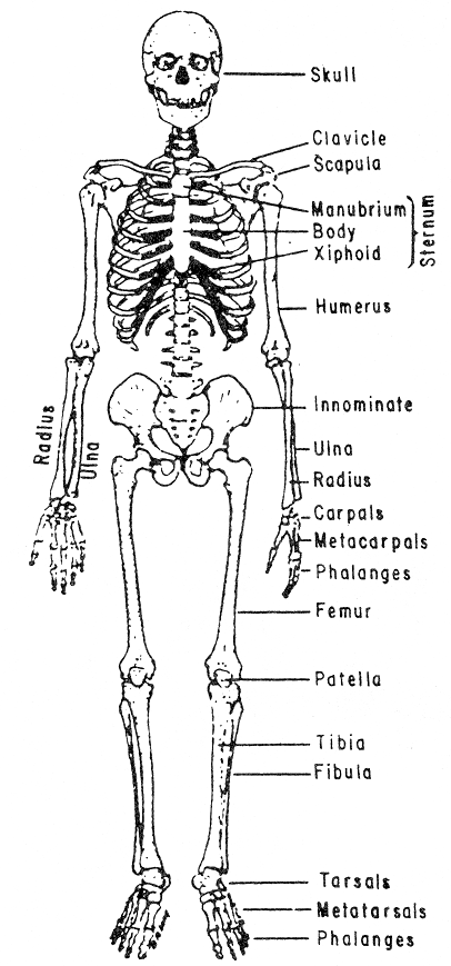
The human skeletal system consists of two primary divisions: the axial skeleton and the appendicular skeleton. Axial skeleton: The axial skeleton is composed of the skull, vertebral column, and rib cage.It offers a safeguard and shelter for the crucial organs of the body. The skull serves to protect the brain and provide support to the face.
Parts, Functions, Diagram, & Facts Biology Diagrams
Learn about the bones, joints, and skeletal anatomy of the human body with our interactive 3D models. Explore the axial and appendicular skeleton, the skull, the vertebrae, the ribs, the pectoral and pelvic girdles, and the upper and lower limbs.

human skeleton, the internal skeleton that serves as a framework for the body. This framework consists of many individual bones and cartilages.There also are bands of fibrous connective tissue—the ligaments and the tendons—in intimate relationship with the parts of the skeleton. This article is concerned primarily with the gross structure and the function of the skeleton of the normal

Definition, Function and Parts Biology Diagrams
Learn about the structure, function, and types of bones, ligaments, and cartilage that make up the human skeleton. Find out how the skeleton protects vital organs, produces blood cells, and moves the body.